
Alzheimer’s Research: Infusion Of Young Blood Can Restore Mental Capabilities In Old Mice


A new study by researchers at the Stanford University School of Medicine has found that some constituent or agent in the blood of young mice when infused to the bloodsteams of old mice has the ability to restore deteriorating mental capabilities.
The scientists suggest that if the same phenomenon applies to humans, it could initiate a new paradigm for recharging our aging brains, and potentially result in the development of new therapeutic approaches for treating dementias such as Alzheimer’s disease.
In the study, published online May 4 in Nature Medicine, entitled “Young blood reverses age-related impairments in cognitive function and synaptic plasticity in mice“ (published online 4 May 2014; doi:10.1038/nm.3569a) the researchers used sophisticated techniques to investigate numerous important molecular, neuroanatomical, and neurophysiological changes in the brains of old mice that shared the blood of young mice.
The study is coauthored by Saul A Villeda, Kristopher E. Plambeck, Jinte Middeldorp, Joseph M. Castellano, Kira I. Mosher, Jian Luo, Lucas K. Smith, Gregor Bieri, Karin Lin, Daniela Berdnik, Rafael Wabl, Joe Udeochu, Elizabeth G Wheatley, Bende Zou, Danielle A. Simmons, Xinmin S. Xie, Frank M. Longo and Tony Wyss-Coray, of various Stanford U. and UCSF departments, programs and faculties, The Eli and Edythe Broad Center for Regeneration Medicine and Stem Cell Research at San Francisco, the AfaSci Research Laboratory at Redwood City, California, and the Center for Tissue Regeneration, Repair and Restoration, VA Palo Alto Health Care System at Palo Alto, California.
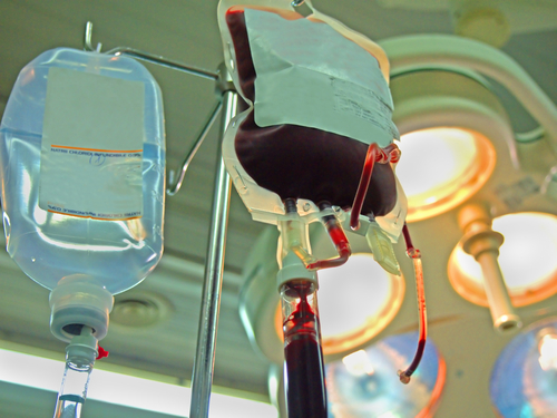
The researchers observe that as human lifespan increases, a greater proportion of the population is suffering from age-related cognitive impairments, making it important to develop means of combating these effects. In the study they report that exposure of an aged animal to young blood can counteract and reverse pre-existing effects of brain aging at the molecular, structural, functional and cognitive level.
The coauthors note that at the cognitive level, systemic administration of young blood plasma into aged mice was observed to improve age-related cognitive impairment in both contextual fear conditioning and spatial learning and memory. They note that structural and cognitive enhancements elicited by exposure to young blood are mediated, in part, by activation of the cyclic AMP response element binding protein (Creb) in the aged hippocampus.
[adrotate group=”3″]
Moreover, they report that their data indicate exposure of aged mice to young blood late in life to be capable of rejuvenating synaptic plasticity and improving cognitive function.
The scientists observe that aging drives cognitive impairment and susceptibility to degenerative disorders in healthy individuals by structurally and functionally changing the adult brain. Considering the increase in proportion of the elderly demographic, they cite the importance of identifying means for maintaining cognitive integrity by protecting against, or even counteracting, the aging process, noting that in aged animals, exposure to young blood through heterochronic parabiosis improves stem cell function in muscle, liver, spinal cord, and the brain, and ameliorates cardiac hypertrophy. However, whether enhancements of young blood extend beyond regeneration in the aged brain is as yet undetermined, raising the question of whether young blood can counteract aging and rejuvenate cognitive processes.
 The researchers also conducted a critical experiment that was far from sophisticated, notes Tony Wyss-Coray, PhD, senior author of the study and a professor of neurology and neurological sciences at Stanford comments in a release. Dr. Wyss-Coray notes that the scientists simply compared performance of older mice in standard laboratory tests of spatial memory after these mice had received infusions of plasma (the cell-free part of blood) from young versus old mice, or no plasma at all.
“This could have been done 20 years ago,” says Dr. Wyss-Coray, who is also senior research career scientist at the Veterans Affairs Palo Alto Health Care System. “You don’t need to know anything about how the brain works. You just give an old mouse young blood and see if the animal is smarter than before. It’s just that nobody did it.”
The Wyss-Coray lab at Stanford studies immune and injury responses in aging and neurodegeneration using mouse genetics, behavior, cell culture, and proteomic approaches to try to test this hypothesis. Dr. Wyss-Coray has also co-founded a biotechnology company, Alkahest, to explore the therapeutic implications of the new study’s findings, and serves as director of Alkahests scientific advisory board.
The study’s lead author, Saul Villeda, PhD, now has an active lab of his own as a faculty fellow in anatomy at the University of California-San Francisco. Dr. Villeda was a graduate student at Stanford and, briefly, a postdoctoral scholar under Dr. Wyss-Coray’s direction when the bulk of the work on the Nature Medicine study was performed. Dr. Villeda joined UCSF in 2012 as a UCSF Faculty Fellow in the Department of Anatomy and the Eli and Edythe Broad Center of Regeneration Medicine and Stem Cell Research.
Research focus at the UCSF Villeda lab is on understanding what drives regenerative and cognitive impairments in the aging brain, and moreover how the effects of aging can be reversed in the old brain. The lab concentrates on three areas. First, looking at how immune-related changes in old blood contribute to impairments in neural stem cell function and associated cognitive functions. Second, studying the contribution of the innate immune system to age-related impairments in synaptic plasticity and cognitive function, and Third, looking at how exposure to young blood rejuvenates neural stem cell function, synaptic plasticity and cognitive function in the old brain. Ultimately, the scientists’ goal is to elucidate cellular and molecular mechanisms that promote brain rejuvenation as a means by which to combat age-related neurodegeneration and cognitive dysfunction.
 “We’ve shown that at least some age-related impairments in brain function are reversible. They’re not final, Dr. Villeda says. Previous experiments by Drs. Wyss-Coray, Villeda and colleagues, described in a paper published in 2011 in Nature, had revealed that key regions in the brains of old mice exposed to blood from young mice produced more new nerve cells than did the brains of old mice similarly exposed to blood from old mice. Conversely, exposing young mice to blood from old mice had the opposite effect with respect to new nerve-cell production, and also reduced the young mice’s ability to navigate their environments.
However that earlier work didn’t directly assess the impact of young mouse blood on older mouse behavior. In this latest study, researchers checked both for changes within nerve circuits and individual nerve cells and for demonstrable improvements in learning and memory — first examining pairs of mice whose circulatory systems had been surgically conjoined, since members of such pairs, known as parabiotic mice, share a pooled blood supply.
Dr. Wyss-Coray’s group paid special attention in these parabiotic mice the hippocampus area of the brain, which in both mice and humans is critical to the formation of certain types of memories, notably recollection and recognition of spatial patterns. “That’s what you need to use when, for example, you try to find your car in a parking lot or navigate around a city without using your GPS system,” Dr. Wyss-Coray explains.
Experience alters hippocampal activity and anatomy, with studies having found, for instance, that a veteran London cabdrivers’ hippocampus is larger than it was when the driver was first hired, and larger than the average person’s. The hippocampus is also extremely vulnerable to the normal aging process, showing early erosion in function as people grow older. In dementias such as Alzheimer’s disease, this hippocampal deterioration is accelerated, leading to an inability to form new memories.
“We know that detrimental anatomical and functional changes occur in the hippocampus as mice and people get older,” says Dr. Villeda. “This is just from natural aging. We’re all heading in that direction.”
When the investigators compared hippocampi from old mice whose circulatory systems had been conjoined with those of young mice to hippocampi from old mice that had been paired with other old mice, they found consistent differences in several biochemical, anatomical and electrophysiological measures known to be important to nerve-cell circuits encoding of new experiences for retention in the cerebral cortex. Dr. Wyss-Coray and his collaborators are working to discover which specific factors in the blood of young mice can recharge the brain of an old mouse.
They observe that the hippocampi of older mice that had been conjoined to younger mice more closely resembled those of younger mice than did the hippocampi of older mice similarly paired with old mice. The old mice paired with young mice made greater amounts of certain substances that hippocampal cells are known to produce when learning is taking place, they cite as an example, noting that hippocampal nerve cells from older members of old-young parabiotic pairs also showed enhanced ability to strengthen connections between one nerve cell and another essential to learning and memory. “It was as if these old brains were recharged by young blood,” Dr. Wyss-Coray observes.
Drs. Villeda, Wyss-Coray and their associates next subjected regular older mice to a test in which the mice were trained to quickly locate a submerged platform in a water-filled container. The mice had to speedily orient themselves using memory cues provided by their surroundings. The investigators injected old mice intravenously with plasma from young or old mice and ran them through the test. Typically, untreated older mice did poorly compared to young mice, as they did when injected with plasma from old mice. However, if they were infused with young mice’s plasma they did much better. Likewise with results of another test in which mice were trained to freeze in fear when plunked into a particular environment. The better they recognized that environment, the longer they would freeze. Older mice typically freeze for a shorter period of time than younger ones do. Again, freezing times for older mice given young plasma, but not old plasma, increased significantly.
The scientists also observed that in both tests, improvement vanished if the plasma provided to the old mice had first been subjected to high temperatures. Heat treatment can denature proteins, so they suggest this hints that a blood-borne protein, or group of them, may be responsible for the cognitive improvements seen in old mice given young mouse plasma.
“There are factors present in blood from young mice that can recharge an old mouses brain so that it functions more like a younger one,” Dr. Wyss-Coray notes. “We’re working intensively to find out what those factors might be and from exactly which tissues they originate. “We don’t know yet if this will work in humans,” he continues, adding that he hopes to find out sooner rather than later. A near-term goal of Dr. Wyss-Coray’s company is to test this proposition through a clinical trial.
A report by The New Scientist’s Andy Coghlan notes that two studies from Dr. Amy Wagers’s lab at Harvard University’s Stem Cell Institute, published in the journal Science ( Science, DOI: 10.1126/science.1251141; Science, DOI: 10.1126/science.1251152) have also found that a blood protein called growth differentiation factor 11 (GDF11) can repair age-related damage in the brains and muscles of old mice, returning them to a more youthful state, noting that if this effect would also work in humans, it could have huge potential for treating a wide-range of age-related diseases.
One of the Science studies explored the potential of GDF11 in muscle regeneration and the other in brain regeneration. Coghlan reports that in the brain study, when researchers injected 15-month-old mice (approximately mid-way through their natural life span) with GDF11 daily for a month, blood vessel volumes in their brains increased by 50 percent and the number of brain stem cells by 29 percent — both factors being known to improve brain function. Muscle fibers in old mice injected with GDF11 also doubled in size to match that of two-month old mice, and the article notes that images from an electron microscope show a striking reordering of muscle fibres from a disordered state to the highly regular appearance of young muscle. Physical endurance of the mice also improved, enabling them to spend an average of 57 minutes on a treadmill compared with 35 minutes for untreated old mice.
For a commentary on the Nature Medicine study entitled “Young blood rejuvenates old brains,” by Steven M Paul & Kiran Reddy, see: http://web.stanford.edu/group/twclab/cgi-bin/images/TWClab/publications/2014_villeda_natmed_newsviews.pdf
Other Stanford co-authors of the Nature Medicine paper were Frank Longo, MD, PhD, professor and chair of neurology and neurological sciences; postdoctoral scholars Jinte Middeldorp, PhD, and Joseph Castellano, PhD; graduate students Kira Mosher and Gregor Bieri; research associates Daniela Berdnik, PhD, and Rafael Wabl; senior research scientist Danielle Simmons, PhD; and senior scientist Jian Luo, MD, PhD.
The study was funded by the U.S. Department of Veteran Affairs., the California Institute for Regenerative Medicine and the National Institute of Aging (grants AG045034 and AG03144).
Information about Stanford’s Department of Neurology and Neurological Sciences, which also supported this work, is available at http://neurology.stanford.edu/
Sources:
Stanford University
University of California at San Francisco
Harvard University Stem Cell Institute
Nature Medicine
Science
Image Credits:
Stanford University
University of California at San Francisco
Harvard University Stem Cell Institute