
Amyloidosis Links Type 2 Diabetes and Alzheimer’s Disease


 Researchers at the Uppsala University in Sweden recently found a molecular link between Type 2 Diabetes and Alzheimer’s disease based on a rare condition called amyloidosis that occurs in both conditions. The study is published in the American Journal of Pathology.
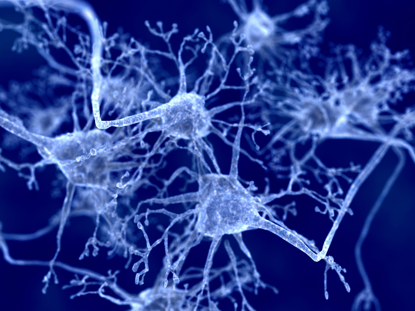
Amyloidosis results from the accumulation of inappropriately folded proteins called amyloids. When proteins that are normally soluble in water fold to become amyloids, they become insoluble and deposit in tissues or organs, disrupting normal function.
In their study titled “In Vivo Seeding and Cross-Seeding of Localized Amyloidosis: A Molecular Link between Type 2 Diabetes and Alzheimer Disease” Gunilla T. Westermark, Ph.D., Department of Medical Cell Biology, Uppsala University, Sweden and colleagues discovered that brain amyloid can motivate the growth of fibrils in the murine pancreas. This pancreatic amyloid can be found in human senile brains.
Patients with Type 2 diabetes commonly have Islet amyloid, a polypeptide (IAPP), derived from its precursor proIAPP. Evidence has shown that accumulation of IAPP can lead to beta-cell death. In Alzheimer’s disease, evidence has shown beta-amyloid deposits in the brain cortex and blood vessels.
“Several soluble proteins are amyloid forming in humans. Independent of protein origin, the fibrils produced are morphologically similar,” said Gunilla T. Westermark, PhD, Department of Medical Cell Biology at Uppsala University in a recent news release. “There is a potential for structures with amyloid-seeding ability to induce both homologous and heterologous fibril growth. Heterologous seeding between IAPP and beta-amyloid may represent a molecular link between AD and T2D.”
Recent evidence shows that patients with Type 2 diabetes are two times more likely to develop Alzheimer’s disease. Results from this study indicate that amyloidosis may link the two conditions.
In their experiments, the researchers injected transgenic mice expressing human IAPP with fibrils of synthetic IAPP, proIAPP, or beta-amyloid after ten months under a high calorie diet the researchers analyzed the tissue. Compared to all three fibrils, the islets with amyloid were increased. There were no deposits of amyloid in the spleen, kidney, liver, heart, or lungs.
In the next experiment, the researchers examined pancreatic and brain tissues. Based on an antibody method, the results showed that sections of the pancreas with islet amyloid from patients with Type 2 diabetes had no beta-amyloid immunoreactivity, while all other samples were found to be immunoreactive for IAPP.
In other experiments, the researchers tested if IAPP and beta-amyloid are present in the human brain tissue. Tissue samples were derived from the temporal cortex of patients with Alzheimer’s disease and a group of patients with frontotemporal dementia, progressive supranuclear palsy (PSP), or no neurological diagnosis. Results revealed IAPP reactivity in all samples, and patients with Alzheimer’s disease were found to have 1.4-times higher IAPP concentrations than samples from non-AD patients.
“It is not clear if IAPP found in brain is locally produced or derived from pancreatic beta-cells,” commented Dr. Westermark in the news release. “Cross-seeding by other amyloid aggregates or perhaps by other types of aggregates offers one possible mechanism for initiation of amyloid formation. Interactions between amyloid and other aggregation-prone proteins may be of great importance in the development of protein-misfolding diseases.”