Alzheimer’s Proteins In Brain Tissue Extracted More Easily by New Technique

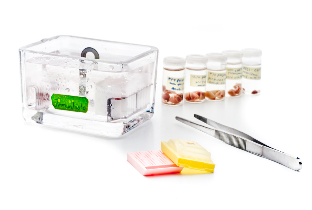
Researchers from NYU Langone Medical Center developed a novel strategy to extract large quantities of brain protein from formalin-fixed, paraffin-embedded (FFPE) tissues in patients with Alzheimer’s, an advancement that may lead to a new understanding of brain protein changes found in the disease. The study entitled “Proteomic analysis of neurons microdissected from formalin-fixed, paraffin-embedded Alzheimer’s disease brain tissue” was published in Scientific Reports.
Neurodegenerative brain disorders like Alzheimer’s, which affect an estimated 5.3 million people — mostly older women — in the U.S., share some common symptoms, like recent-event memory loss, poor judgment, confusion, and changes in mood and personality.
Though advances in science have led to progress in understanding some aspects related to Alzheimer’s, the disease’s exact causes and underlying mechanisms are not fully known. Evidence to date points to several risk factors that contribute toward Alzheimer’s development, including genetics, infections with herpes simplex virus, and decreased synthesis of the neurotransmitter acetylcholine. Other studies highlight the presence of abnormal levels of extracellular amyloid beta (Aβ) plaque deposits and tau protein in brain tissue. These folded proteins are generally insoluble, which makes them difficult to study using FFPE tissue, or tissue saturated in wax and cut for analysis that is widely available for medical research.
In this study, researchers used a tool called laser-capture micro dissection to extract more than 400 proteins — from neurons in the brain region known as the temporal cortex — of 10-year-old archival FFPE tissue with Alzheimer’s disease. Formic acid was used to dissolve the tissue in order to collect the proteins in a larger number than ever previously extracted.
“This new technique allows for more precise examination of formalin-preserved tissue from hundreds of brain banks around the world, and holds particular value in studying the pathogenesis of Alzheimer’s disease and other neurodegenertative diseases, particularly for examining”, said the study’s senior investigator, Thomas Wisniewski, MD, director of the NYU Langone Center for Cognitive Neurology, and the Lulu P. and David J. Levidow Professor of Neurology, and professor of pathology and psychiatry at NYU School of Medicine. “Previously these types of studies required frozen tissue.”
“Formalin-fixed paraffin-embedded tissue is plentiful,” Dr. Wisniewski added, emphasizing the importance of his team’s work. “We think that the technique is relatively simple and reproducible, and offers a highly sensitive method to detect proteins. It will offer researchers an important new tool for examining proteins using very small amounts of archived tissue.”


