
New MRI Method May Help Detect Early Stage Alzheimer’s
Written by |
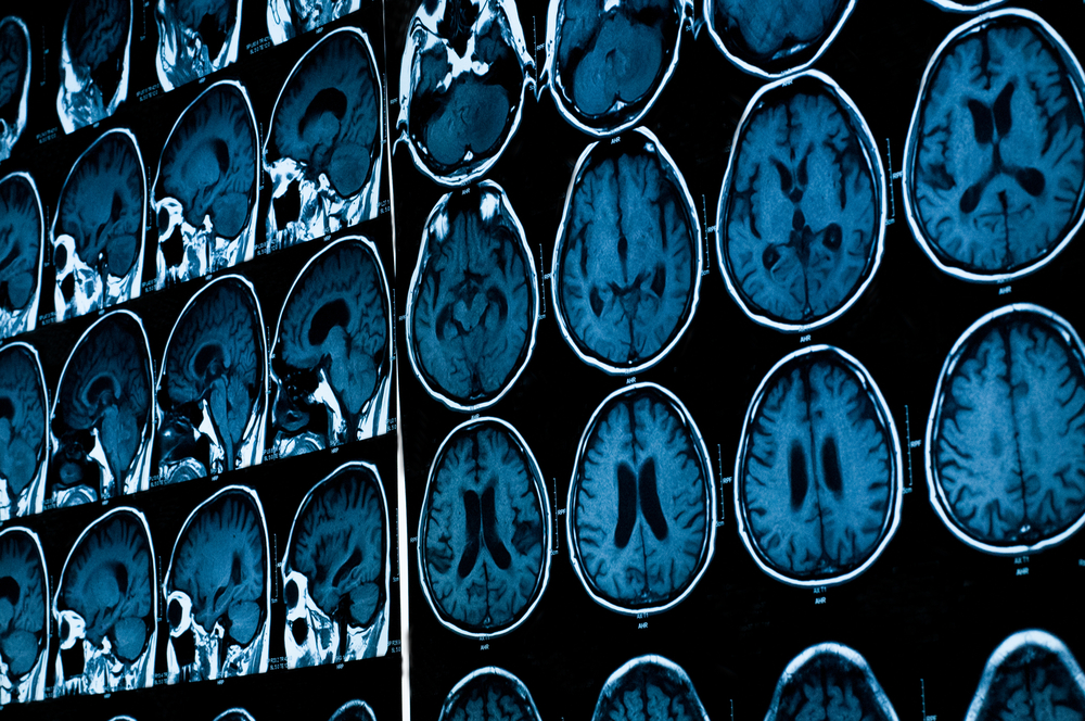


 A new, sensitive molecular magnetic resonance imaging contrast probe was recently developed by a team of researchers from Northwestern University. The study, entitled “Towards non-invasive diagnostic imaging of early-stage Alzheimer’s disease” was published in Nature Nanotechnology by Kirsten L. Viola, first author of the study, and led by Dr. William L. Klein, from the Department of Neurobiology, Northwestern University, Evanston, Illinois, USA, and Dr. Vinayak P. Dravid, from the Department of Materials Science and Engineering, Northwestern University, Evanston, Illinois, along with colleagues.
Alzheimer’s disease is the most common form of dementia, characterized by two important pathological features: amyloid-β plaques and neurofibrillary tangles. The amyloid hypothesis of Alzheimer’s disease suggests that the excessive accumulation of amyloid-β peptide leads to neurofibrillary tangles composed of aggregated tau hyperphosphorylated. It has been postulated that early-stage biomarkers that induce memory loss are composed of Aβ oligomers. In early stages of Alzheimer’s disease, the amyloid beta oligomers in the brain attack synapses of neurons, affecting and impairing memory and, ultimately, leading to the death of neurons. With the development of the disease, the amyloid betas begin to accumulate and attach to each other, inducing the formation of amyloid plaques. However, oligomers are the early intruders — they appear up to one decade earlier than the plaques.
The imaging technique, called positron emission tomography, has been used to detect the pathological features of Alzheimer’s disease using probes that bind amyloid fibrils that was usually performed at a late stage of Alzheimer’s disease when therapeutic therapy was already not helpful. However, it seems that amyloid fibrils are not directly connected to the triggering of the disease, but instead the oligomers are the early players in the development of the disease. Importantly, the amyloid beta oligomers seem to be the biomarkers for the onset of Alzheimer’s disease and proceeding memory loss.
The research team reported a sensitive molecular magnetic resonance imaging contrast probe that consists of oligomer-specific antibodies bounded to magnetic nano structures that specifically target Aβ oligomers on cells and brain tissues to generate a magnetic resonance imaging signal. The researchers tested these probes intranasally in an Alzheimer’s disease mouse model and observed that they immediately reached the hippocampal Aβ oligomers. Moreover, the authors isolated samples of human brain tissue and performed magnetic resonance imaging measurements, and found a signal that enabled to distinguish Alzheimer’s patient’s samples from controls. Notably, the nanostructures that bind neurotoxic Aβ oligomers may be important for assessing the efficacy of novel drugs and ultimately for early-stage Alzheimer’s disease diagnosis and disease management.
“Non-invasive imaging by MRI of amyloid beta oligomers is a giant step forward towards diagnosis of this debilitating disease in its earliest form,” said Dr. Dravid, the Abraham Harris Professor of Materials Science and Engineering at the McCormick School of Engineering and Applied Science, in a press release. “This MRI method could be used to determine how well a new drug is working. If a drug is effective, you would expect the amyloid beta signal to go down,” added Dr. Dravid.
Leave a comment
Fill in the required fields to post. Your email address will not be published.