Novel Culture System Replicates Alzheimer’s Disease Development; Confirms Amyloid Hypothesis; Should Significantly Reduce Drug Development Time/Cost

An innovative laboratory culture system developed by an international team of investigators led by scientists of the Genetics and Aging Research Unit at Massachusetts General Hospital (MGH) provides the first clear evidence supporting the hypothesis that deposition of beta-amyloid plaques in the brain is the first step in a cascade of events underlying development of Alzheimer’s disease (AD). Using a novel three-dimensional culture system, the Mass. General scientists have succeeded, for the first time, in reproducing the full course of the devastating neurodegenerative disease’s development, and also identify the essential role in that process of a particular enzyme, the inhibition of which could be a therapeutic target.
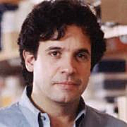
 “Originally put forth in the mid-1980s, the amyloid hypothesis maintained that beta-amyloid deposits in the brain set off all subsequent events the neurofibrillary tangles that choke the insides of neurons, neuronal cell death, and inflammation leading to a vicious cycle of massive cell death,” says Rudolph Tanzi, PhD, director of the MGH Genetics and Aging Research Unit and co-senior author of the report receiving advance online publication in the journal Nature in a Mass. General release. “One of the biggest questions since then has been whether beta-amyloid actually triggers the formation of the tangles that kill neurons. In this new system that we call Alzheimers-in-a-dish, we’ve been able to show for the first time that amyloid deposition is sufficient to lead to tangles and subsequent cell death.”
The Nature paper entitled “A three-dimensional human neural cell culture model of Alzheimer’s disease” (Published online 12 October 2014 Nature (2014) doi:10.1038/nature13800), is coauthored by Se Hoon Choi, Young Hye Kim, Matthias Hebisch, Christopher Sliwinski, Carla DAvanzo, Hechao Chen, Basavaraj Hooli, Caroline Asselin, Justin B. Klee, Can Zhang, Dora M. Kovacs, Rudolph E. Tanzi, and Doo Yeon Kim of the Genetics and Aging Research Unit, MassGeneral Institute for Neurodegenerative Disease, Massachusetts General Hospital, Harvard Medical School, Charlestown, Mass.; Young Hye Kim of the Division of Mass Spectrometry Research, Korea Basic Science Institute, Cheongju-si, Chungbuk, South Korea; Matthias Hebisch and Michael Peitz of the Institute of Reconstructive Neurobiology, Life and Brain Center, University of Bonn and Hertie Foundation, Bonn, Germany; Seungkyu Lee, Brian J. Wainger, and Clifford J. Woolf of the FM Kirby Neurobiology Center, Boston Childrens Hospital and Harvard Stem Cell Institute, Boston; Julien Muffat of the Whitehead Institute for Biomedical Research, Cambridge, Massachusetts; and Steven L. Wagner of the Department of Neurosciences, University of California, San Diego, La Jolla, California.
The coauthors note that Alzheimer s disease is the most common form of dementia, characterized by two pathological hallmarks: amyloid plaques and neurofibrillary tangles. The amyloid hypothesis of Alzheimer’s disease posits that the excessive accumulation of amyloid-peptide leads to neurofibrillary tangles, but to date, no single disease model had serially linked these two pathological events using human neuronal cells.
The researchers observed that while Mouse models with the inherited early-onset form of familial Alzheimer’s disease (FAD) mutations exhibit amyloid-induced synaptic and memory deficits, they do not fully recapitulate other key pathological events of Alzheimer’s disease, including distinct neurofibrillary tangle pathology. Other models succeed in producing tangles but not plaques. Cultured neurons from human patients with Alzheimer’s exhibit elevated levels of the toxic form of amyloid found in plaques and the abnormal version of the tau protein that makes up tangles, but not actual plaques and tangles.
 The Mass. General release notes that Genetics and Aging Research Unit investigator Doo Yeon Kim, PhD, co-senior author of the Nature paper, realized that the liquid two-dimensional systems usually used to grow cultured cells poorly represent the gelatinous three-dimensional environment within the brain. Instead the MGH team used a gel-based, three-dimensional culture system to grow human neural stem cells that carried variants in two genes the amyloid precursor protein and presenilin 1 known to underlie early-onset familial Alzheimer’s Disease (FAD). Both of those genes were co-discovered in Dr. Tanzi’s laboratory.
After growing for six weeks, the FAD-variant cells were found to have significant increases in both the typical form of beta-amyloid and the toxic form associated with Alzheimer’s. The variant cells also contained the neurofibrillary tangles that choke the inside of nerve cells causing cell death. Blocking steps known to be essential for the formation of amyloid plaques also prevented the formation of the tangles, confirming amyloids role in initiating the process. The version of tau found in tangles is characterized by the presence of excess phosphate molecules, and when the team investigated possible ways of blocking tau production, they found that inhibiting the action of an enzyme called GSK3-beta known to phosphorylate tau in human neurons prevented the formation of tau aggregates and tangles even in the presence of abundant beta-amyloid and amyloid plaques
“This new system, which can be adapted to other neurodegenerative disorders, should revolutionize drug discovery in terms of speed, costs and physiologic relevance to disease,” says Dr. Tanzi. Testing drugs in mouse models that typically have brain deposits of either plaques or tangles, but not both, takes more than a year and is very costly. With our three-dimensional model that recapitulates both plaques and tangles, we now can screen hundreds of thousands of drugs in a matter of months without using animals in a system that is considerably more relevant to the events occurring in the brains of Alzheimer’s patients.”
At Mass. General’s Genetics and Aging Research Unit’s Tanzi Lab, Dr. Tanzi and his team conduct genome-wide association screens to identify novel genes associated with AD and autism spectrum disorders. Since 1982, he has focused his studies on Alzheimer’s disease, and isolated the first familial Alzheimer’s disease (FAD) gene, known as the amyloid beta-protein (A4) precursor (APP) in 1987, as well as another in 1995, called presenilin 2. He also collaborated on the isolation of the second FAD gene, presenilin 1. In 1993, Dr. Tanzi isolated the gene responsible for the neurological disorder known as Wilson’s disease, and over the past 25 years, he has collaborated on studies identifying several other neurodegenerative disease genes including those causing amyotrophic lateral sclerosis and neurofibromatosis.
Dr. Tanzi’s laboratory first discovered that the metals zinc and copper are necessary for the formation of neurotoxic assemblies of the AD-associated peptide, A-beta, the main component of beta-amyloid deposits in brains of AD patients. These studies have led to ongoing clinical trials for treating and preventing AD by targeting A-beta metal interactions. Dr. Tanzi was also involved in the first studies implicating gamma-secretase modulators as therapeutics for AD. He is currently carrying out genome wide association screens to identify novel genes associated with AD and autism spectrum disorders. Candidate disease genes are then characterized at the molecular, cell biological, and biochemical levels to elucidate disease mechanisms.
“My research is aimed at identifying and characterizing Alzheimer’s disease-associated gene mutations and variants with the ultimate goal of defining the molecular, cellular, and biochemical events leading to neuronal cell death in the brains of AD victims,” Dr. Tanzi notes. “In addition to identifying risk factors for Alzheimer’s disease, we address the mechanisms underlying the etiology and pathogenicity of the genes responsible for Alzheimer’s disease through the application of molecular, cell biological, and biochemical strategies. Our ongoing research in AD and autism follows a basic roadmap, which includes disease gene discovery, translational and functional studies to identify pathogenic gene variants and mutations, molecular biological and biochemical studies to elucidate pathways that have been impacted by disease-associated gene changes, and novel drug screening assays to identify small molecules or supplements that can halt or reverse pathogenic molecular and biochemical phenotypes at the cellular level.”
“A significant portion of AD involves inheritance of defective genes variants and mutations that influence ones lifetime risk for the disease,” he continues. “To date, four different genes have been firmly implicated to play a role in familial Alzheimer’s disease (FAD). Our laboratory has co-discovered three of these genes: the amyloid protein precursor [APP], presenilin 1 [PSEN1], and presenilin 2 [PSEN2]) that harbor defects causing “early-onset” forms of the disease with 100 percent certainty under 60 years old. Our current studies are targeted toward determining the pathogenic mechanisms by which these gene defects contribute to the neurodegenerative process in the brains of AD patients. The known late-onset AD gene, APOE, accounts for 30-40% of the genetic variance of AD. Thus, we have been carrying out genome-wide association scans to search for novel AD genes using >1300 AD families 1M SNP chips. We have also collaborated on the completion of a genome-wide association screen of families with autism (N=3700) and identified novel candidate genes on chromosomes 5 and 16. Our research in both AD and autism follows a basic road map starting with disease gene discovery, then proceeding into translational and functional studies to identify pathogenic gene variants and mutations, and finally progressing to novel drug screening to identify small molecules that can halt or reverse pathogenic molecular and biochemical phenotypes at the cellular level.”
Dr. Tanzi is the Kennedy Professor of Child Neurology and Mental Retardation, and Dr. Kim is an assistant professor of Neurology at Harvard Medical School. Se Hoon Choi, PhD, and Young Hye Kim of the MGH Genetics and Aging Research Unit are co-lead authors of the Nature paper. The study was supported by a grant from the Cure Alzheimers Fund and by National Institute of Health grants 5P01AG15379 and 5R37MH060009.
Sources:
Massachusetts General Hospital
Nature
Image Credits:
Massachusetts General Hospital


