
Watching Proteins Unfold: New Insights Could Impact Alzheimer’s
Written by |


Scientists at the University of Illinois have created an exciting new method for examining how proteins change their shape right inside cells, using fluorescent markers and a specialized microscope. They recently published their work in the December 1st issue of the journal Public Library of Science One (PLoS One).
Many neurological diseases are characterized, and possibly caused by mis-folded proteins that muck up the cells of the brain and kill them. Examples of diseases and associated aberrant proteins include: beta-amyloid protein in Alzheimer’s disease, huntingtin protein in Huntington’s disease and alpha-synuclein in Parkinson’s disease. Understanding how proteins in these diseases fold, mis-fold and how they are carried through cells and in the body could greatly help with understanding and possibly even treating these conditions.
This is where the technology developed by Chemistry professor Martin Gruebele and his graduate students Minghao Guo and Hannah Gelman may help. For example, tracking protein transport and folding in Alzheimer’s disease animal models may assist with understanding how the disease progresses. In the past, scientists simply studied the remnants of beta-amyloid protein in the already degenerated Alzheimer’s diseased brain, making assumptions of how beta-amyloid deposits developed and killed cells.
Gruebele pointed out that with the technology developed by his group: “We’re looking at the earliest stages of disease, the initial phases of transport of bad proteins…..we think the fibrils are just an end product that’s left over when the cell dies, and the actual killing mechanism has to do with migration of the protein to specific places in the cell like the outer membrane. Understanding how these mechanisms work at a fundamental level is going to give people more handles on where to look to cure things.”
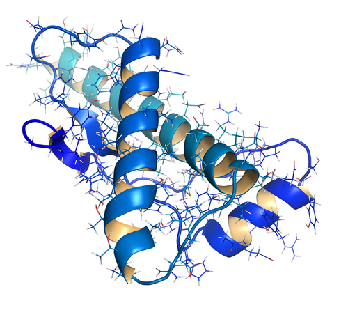
As an example of how protein folding could go wrong, the researchers studied a mutant form of the enzyme phosphoglycerate kinase (PGK) and compared it to the stable form of the same protein. PGK’s normal function is to break down glucose (sugar) in the body to be used as energy. They measured how the protein’s motion slows down when it unfolds using a fluorescence microscope, then used 3-D modeling to estimate the motion of protein folding over time. According to their analysis, when the mutant protein began to unfold within the cell, the scientists noticed that it began “sticking” to the cytosol–the inside of the cell. It also moved more slowly than the normal protein. A similar process might be going on in neurological diseases.
In their study they concluded, “The unfolded protein clearly diffuses more slowly than the folded protein, whether due to its larger size, ‘sticking’, or both.”
Greater understanding of the ongoing processes that control proteins might open up exciting new doors to understanding and treatment of diseases that involve abnormally folded proteins.


Leave a comment
Fill in the required fields to post. Your email address will not be published.